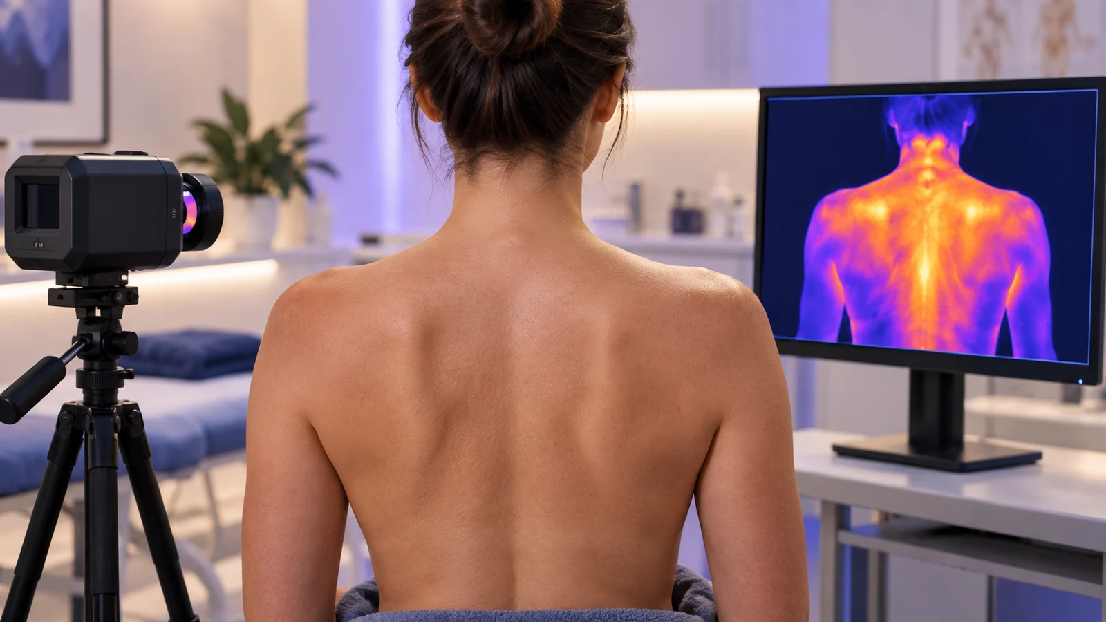
A thermal camera does not see pain. It does not see inflammation, a torn ligament or a compressed nerve. It sees the one thing your skin does in response to all of those, and it sees that one thing extraordinarily well.
Understanding what that one thing is turns a colorful picture into clinical information. Misunderstanding it produces the two classic errors of thermography: reading a warm patch as proof of injury, and reading an unremarkable image as proof of health.
This is a guide to what the camera actually measures, why clinicians compare one side of the body with the other, what a thermatome is, and where a thermogram's knowledge stops.
The Camera Measures Heat Leaving the Skin
Every object warmer than absolute zero radiates infrared energy. A medical thermal camera measures the long-wave infrared radiation, roughly 8 to 14 micrometers in wavelength, leaving the skin surface, and converts it into a temperature for each pixel. Modern medical cameras resolve differences of a few hundredths of a degree.
The word that matters in that sentence is surface. The sensor receives radiation from the outermost layer of skin and nothing else. A joint, a muscle or a nerve two centimeters down never appears in the image directly. Deep structures influence a thermogram only by changing the temperature of the skin above or near them.
So what sets skin temperature? Three things. Heat conducted from tissues beneath, such as a working muscle or an inflamed joint. The environment, which is why acquisition happens in a controlled room after an acclimatization period. And, above all, blood flow through the dermal microcirculation, the network of small vessels a millimeter or two beneath the surface.
That blood flow is governed largely by the sympathetic branch of the autonomic nervous system, which constricts and dilates those vessels continuously and without conscious involvement. Think of the skin as a radiator whose valves are set by a thermostat you cannot feel. The camera reads the radiator. In doing so, it reads the valves.
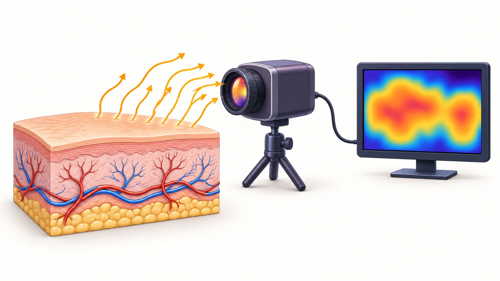
Conceptual illustration: blood flow influences skin temperature. The camera detects infrared radiation from the surface and converts it into a temperature map; it does not image structures beneath the skin.
Why Clinicians Compare Left With Right
Absolute skin temperature is an unreliable witness. It changes with the room, the time of day, caffeine, hydration, exercise and the individual. A forearm reading of 31.2 °C means very little on its own.
Left-right symmetry is a far more stable reference, because both sides of the body share the same environment, the same circulation and the same autonomic tone. In a study of 39 healthy men, the largest side-to-side temperature difference at any site was 0.4 °C, with a spread of 0.3 °C in whole-body views and 0.15 °C in regional views. By common clinical convention, asymmetries beyond roughly 0.5 to 0.7 °C are treated as a signal worth explaining.
An earlier Johns Hopkins series pointed the same way. Across 40 matched regions in 90 asymptomatic adults, mean asymmetry ranged from 0.18 °C at the forehead to 0.38 °C at the foot, and the values held on repeat measurement over five years.
This side-to-side difference is what clinicians call thermal asymmetry, usually written as ΔT. It is the core measurable output of medical thermography.
The logic is the same as comparing two identical plants in the same window. You do not need to know the room's humidity to notice that one of them is wilting. The body runs this experiment on itself every moment, and the thermal camera is the instrument that reads the result.
Thermatomes: The Skin Is Wired in Segments
Most clinicians know the dermatome chart: the striped map of skin regions, each supplied for sensation by a single spinal nerve root. Less familiar is that the skin's blood vessels are wired in segments too.
The sympathetic fibres that control cutaneous blood flow leave the spinal cord by segment and travel to the skin alongside peripheral nerves. The territory of skin whose vessels are governed by a given nerve is that nerve's thermatome. Thermatome maps overlap with dermatome maps but do not coincide, because the sympathetic and sensory fibres take partly different routes to the surface.
The clearest evidence that skin temperature follows nerve territory comes from anesthesia, where the effect is produced deliberately. In a 2023 prospective study of 80 patients receiving a supraclavicular brachial plexus block, the local anesthetic silenced the sympathetic fibres along with the sensory ones. Vessels in the blocked territories dilated, and the skin warmed in exactly the regions supplied by the ulnar, median and radial nerves.
At 15 minutes, a temperature rise in each nerve's territory excluded a failed block with 100 percent sensitivity and 90 to 94 percent specificity. A similar approach has been used to map the extent of thoracic paravertebral blocks dermatome by dermatome. In both cases the camera is reading which segments of the nervous system have opened their valves.
For a clinician looking at a thermogram, this changes what a pattern means. A cool band that follows a nerve's territory is a different observation from a cool patch that follows the outline of a bruise. Warmth confined to one segment is different from diffuse warmth across a limb. The pattern carries more information than the color.
The Viscerocutaneous Reflex: When Skin Reports on an Organ
Sensory nerves from internal organs enter the spinal cord at the same segments as the sympathetic nerves that leave it for the skin. This convergence is why heart pain is felt in the left arm and gallbladder pain below the right shoulder blade. The brain cannot always tell which input arrived on a shared line.
The same convergence can run outward. Irritation of an organ can alter sympathetic outflow to the skin of the corresponding segment, changing its blood flow and therefore its temperature at a site that may be far from the organ itself. Thermology has described such viscerocutaneous, or viscerosomatic, reflex patterns since the 1980s, mainly in abdominal and pelvic pain.
This section deserves a caution. The evidence here is older, smaller and pattern-based than the nerve-block literature. A segmental skin-temperature change that has no musculoskeletal explanation is a reason to widen the differential and involve the right specialist. It is not, by itself, a finding about any organ.
What a Thermogram Cannot Tell You
Knowing the physiology makes the limits obvious.
- It shows no structure. A tear, a herniated disc or a fracture is invisible to it. Structural imaging remains the tool for structure.
- Warm does not mean inflamed. Recent exercise, sunlight, a topical product, a hot drink or a draft in the room can all warm skin. This is why protocols insist on acclimatization and standard conditions.
- Cool does not mean nerve injury. Vasoconstriction has many causes, from anxiety to a cold hand resting on the thigh a minute earlier.
- One image is one moment. Physiology moves. Repeated imaging over days or weeks is often more informative than a single frame.
- It does not diagnose. Medical thermography is an adjunct to clinical judgment. It provides objective physiological data that supports assessment alongside history, examination and other imaging.
Reading One, in Order
Clinicians trained in thermography tend to read an image in the same sequence, and the sequence follows the physiology above.
- Acquisition discipline first. A controlled room, an acclimatization period, no recent exercise or topical products, standardized positions. Without this, nothing that follows is trustworthy.
- Symmetry. Compare each region with its counterpart on the other side. The ΔT is the primary observation.
- Pattern. Ask what any asymmetry follows: a nerve territory, a joint, a muscle, a vascular distribution, or none of these.
- Context. Place the thermal finding beside the history, the examination and any structural imaging. The thermogram contributes a physiological signal; the clinician decides what it means.
- Time. Where possible, repeat. A trajectory is worth more than a snapshot.
Where Vizbodx Fits
Vizbodx software is built around the same order of operations. It quantifies bilateral thermal asymmetry on anatomically corresponding regions, tracks those regions across sessions so that change over time can be seen, and presents its AI analysis only after the clinician has recorded an independent read, an architecture the company calls Clinician-First AI.
The foundations described in this article are also the starting point of Vizbodx Academy, launched in August 2026 to give clinicians a structured, evidence-based pathway into clinical infrared imaging. Two of the first three episodes of the companion podcast are titled "What Thermography Really Sees" and "Thermal Symmetry, Thermatomes, and the Viscerocutaneous Reflex," because the company's view is that interpretation has to begin with what the camera physically measures.
None of this replaces the clinician. Infrared imaging is an adjunct that adds an objective physiological layer to assessment. Its value depends on a reader who understands what the layer is made of.
The Report Your Skin Has Always Filed
Your skin has been publishing a report on your nervous system's vascular decisions every second of your life. It does so in the language of temperature, organized by segment, and it publishes it twice, once on each side, so that any departure from symmetry stands out.
A thermal camera is simply the first instrument that reads that report in full. A trained clinician is the one who decides what it means.
Recovery begins with discovery. Discovery begins with knowing what you are looking at.
Vizbodx Inc. is developing AI-powered infrared medical imaging technology designed to detect asymmetric thermal patterns in sports medicine, occupational health and musculoskeletal recovery, often before symptoms emerge. Vizbodx software is an adjunct to clinical judgment; it does not diagnose.
Recovery begins with discovery.
→ Read the original LinkedIn post on the Vizbodx Academy launch → Learn more about Vizbodx
References
- Vardasca R, Ring EFJ, Plassmann P, Jones CD. Thermal symmetry of the upper and lower extremities in healthy subjects. Thermology International. 2012;22(2):53-60. https://www.researchgate.net/publication/227860586_Termal_symmetry_of_the_upper_and_lower_extremities_in_healthy_subjects
- Gamal M, Hasanin A, Adly N, et al. Thermal Imaging to Predict Failed Supraclavicular Brachial Plexus Block: A Prospective Observational Study. Local and Regional Anesthesia. 2023;16:71-80. https://pmc.ncbi.nlm.nih.gov/articles/PMC10263017/
- Infrared thermography for assessment of thoracic paravertebral block: a prospective observational study. 2021. https://pmc.ncbi.nlm.nih.gov/articles/PMC8194215/
- Assessment of neuropathic pain in leprosy patients with relapse or treatment failure by infrared thermography: A cross-sectional study. 2021. https://www.ncbi.nlm.nih.gov/pmc/articles/PMC8491942/
- Uematsu S, Edwin DH, Jankel WR, Kozikowski J, Trattner M. Quantification of thermal asymmetry. Part 1: Normal values and reproducibility. Journal of Neurosurgery. 1988;69(4):552-555. https://pubmed.ncbi.nlm.nih.gov/3418388/
- Osmosis. Viscerosomatics and facilitation. https://www.osmosis.org/learn/Viscerosomatics_and_facilitation
- Vizbodx Inc. Vizbodx Inc. Launches Academy to Establish a New Educational Standard for Clinical Infrared Imaging. PR Newswire, August 4, 2026. https://www.prnewswire.com/news-releases/vizbodx-inc-launches-academy-to-establish-a-new-educational-standard-for-clinical-infrared-imaging-302842536.html